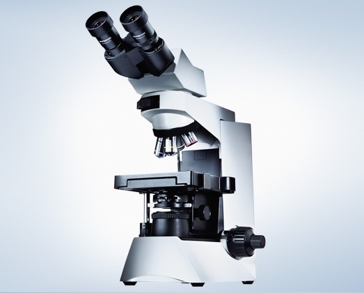
11. The Olympus CX41 Dark-Field Microscope
Specifications
Olympus CX41 is a powerful universal microscope with the modular principle of construction. This microscope is developed for laboratory researches. It has binocular and trinocular drawtubes and provides a zoom range of 20x to 1250x. Its optical system is set to infinity. A built-in illuminant is based on a 30-W halogen bulb light source. There is a possibility of mounting phase-contrast and dark-field condensers, a fluorescence unit, polarizers as well as non-immersion and water-immersion lenses. One can also make photo- and video documentation.
Pentagonal rotary turret of the Olympus CX41 microscope has an additional port for location of polarizer and sample wafers. Any lenses, various types of condensers and observation drawtubes can be included in a complete set of the microscope. The fluorescent brightener integrated into the microscope frame, doesn’t change the total magnification and gives the ability to combine the transmitted-light illumination with the indirect illumination.
The trinocular observation drawtube of the Olympus CX41 microscope is provided with the beam splitter. The light flow capacity of the beam splitter is 50:50. A system of film-type photographic registration, a digital camera, or a video camera can be attached to the trinocular drawtube port.
A phase-contrast mode is the most optimum one during the work with low-contrast compounds. The fast switching from the phase-contrast mode to the bright-field mode or fluorescence is also provided.
A dark-field mode provides the highest contrast when one works with light-tight point objects.
A bright-field mode provides a sharp image at all feasible magnifications; it also provides a control of the illuminator color temperature.
A polarization mode is used in medicine, biology, and material science. In this case, the microscope can be used in combination with compensators.
Live Blood Drop Examination Using the Dark-Field Microscope
Live blood drop examination using the dark-field microscope is an alternative and exclusive approach for health diagnostics. If the human body is not tired, not irritated or not slagged, microbes living in it don’t do harm to it. Cells are capable to coexist "peacefully" together with a large number of germs fighting against each other. However, healing natural forces that maintain the balance in the body are not endless. They may be damaged by stresses and overloads. As a result, the harmony between the acid-base balance, the body salt-and-vitamin content, and the functioning of digestive and purifying organs is being disturbed.
Live blood drop examination can provide information on the body state and predisposition to some diseases. Blood is an internal environment of the body through which the metabolism is performed, oxygen gets into cells, and products of their activity (toxins) are eliminated from cells. Recently scientists have come to a conclusion that more accurate data can be provided through the qualitative rather than quantitative examination of formed elements of blood and plasma.
Live blood drop examination makes it possible to evaluate the condition of erythrocytes, their mobility in plasma, their aggregation, and their sludge (stickiness). Analyzing a condition of thrombocytes (platelets), lymphocytes, and leucocytes, one can evaluate activity of immune system and ability of an organism to self-recovery, and also pathological changes of blood composition that cause development of many diseases. Such examination of blood cells and plasma allows tracing various health deviations and/or predisposition to new diseases.
Live blood drop examination makes it possible to diagnose a wide range of diseases and metabolic status such as lipid exchange, protein metabolism, carbohydrate metabolism, and phosphoric-and-calcium metabolism. In turn, the metabolism depends on the pancreas and liver functioning. Live blood drop examination makes it also possible to make a conclusion about various abnormalities, which can cause anemia, atherosclerosis, gout, and malignant tumors. The immune compensation and intestinal dysbacteriosis can be also diagnosed with use of the live blood drop examination.
The Olympus CX41 Dark-Field Microscope Complete Set
No
|
Description
|
Quantity
|
1.
|
CX41RF-5: Microscope frame having a built-in pentagonal rotary turret with analyzer port, illuminant (6 V, 30 W), field stop, rectangular stage with a right-hand control, and a double-sample stage.
|
1
|
2.
|
UYCP: Power cord.
|
1
|
3.
|
6V30W: Halogen bulb (6 V, 30 W).
|
2
|
4.
|
U-CTR30-2-2: Trinocular observation drawtube with a fixed 50:50 light flow distribution, an eyepiece tubes’ slope angle of 30°, an interpupillary distance adjustment range of 48 – 75 mm, diopters’ adjustment on the left eyepiece tube, and the FN20 visual field width. Applied eyepieces: WHB10x, WHB10x-H.
|
1
|
5.
|
WHB 10X-2: Wide-field eyepiece 10х, FN20. It is applied in the U-CBI30-2 and U-CTR30 with the FN20 visual field width.
|
1
|
6.
|
WHB 10XH-2: Wide-field focusable eyepiece 10х, FN20. It is applied in the U-CBI30-2 and U-CTR30 with the FN20 visual field width.
|
1
|
7.
|
CX-SLC: Abbe condenser equipped with a two-position slider and aperture diaphragm with an aperture scale NA of 1.25.
|
1
|
8.
|
CH2-DS: Dark-field insert for placing on the СH2-FH holder to work with objective lenses having an aperture of no more than NA = 0.65.
|
1
|
9.
|
CH2-FH: Holder for optical filters and inserts with a diameter of 32.5 mm.
|
1
|
10.
|
PLCN10X/0.25: Plan Achromat C objective lens with magnification of 10x, working distance WD of 10.5 mm, and numerical aperture NA of 0.25.
|
1
|
11.
|
PLCN40X/0.65: Plan Achromat C 40х objective lens, WD = 0.6 mm, NA = 0.65.
|
1
|
12.
|
PLCN100XOI/1.25: Plan Achromat C 100х objective lens, WD = 0.13 mm, NA = 0.6 – 1.25.
|
1
|
13.
|
DUST PLUG F.RSM THREAD: Dust plug for socket of the bright-field rotary turret.
|
1
|
14.
|
U-TV1X-2: Direct-image adapter 1х.
|
1
|
15.
|
U-CMAD3: C-mount adapter.
|
1
|
16.
|
U-TV0.5XC-3: Photo/video 0.5х C-mount adapter, FN22 for a 2/3" CCD, extended infrared range.
|
1
|
17.
|
Watec WAT221S: Color video camera,
1/2 ”, digital signal processing DSP, 450 TVL (480 TVL, S VHS), 0.1 lx F1.2, composite video output and S VHS (Y /C), objective lens free, electric shutter speed – up to 1/10 000, manual and automatic white balance, backlight compensation.
|
1
|
18.
|
CS thread adapter to С, Watec (Japan).
|
1
|
19.
|
Stabilized power adapter 220 V / 12 V, 0.25 А, Watec (Japan).
|
1
|
20.
|
018 DUST COV BX2: Dust cover.
|
1
|
21.
|
Slide collection box (for 25 pcs).
|
1
|
22.
|
76 х 26-mm slides for microscopy (thickness of 1.0 mm). The slides are resistant to chemical agents, previously washed, 1 000 pcs.
|
1
|
23.
|
20 х 20-mm cover glasses for microscopy (thickness of 0.17 mm). The glasses are ideally suited for fluorescent microscopy, 1 000 pcs. (100 pcs х 10 boxes).
|
1
|
24.
|
Immersion oil in 50-ml lubricator, n = 1,516.
|
1
|
25.
|
LENS CLEANING TISSUE: 60 x 110-mm napless lens cleaning tissues, 100 pcs.
|
10
|
|